


Stress fractures disproportionately affect elite athletes, military personnel, and inexperienced individuals who abruptly begin vigorous physical training.1 The tibia is the most commonly involved site, and 90% of stress fractures in this bone occur along the distal third of the posteromedial cortex. Anterolateral fractures are relatively uncommon (4.6%), but are at higher risk of delayed healing, nonunion, and progression to complete fracture.2,3 On plain radiographs, nonunion is suggested by the “dreaded black line,” a horizontal cortical fracture caused by bony resorption.2 The tensile forces transmitted through the anterolateral tibia contribute to osteoclast proliferation and suboptimal healing, which is further impaired by a lack of regional musculotendinous support.2,3 We present a case of bilateral anterior mid-tibial stress fractures in the background of primary hyperparathyroidism (PHPT).
Primary hyperparathyroidism is most often caused by a benign adenoma; however, it can be the result of hyperplastic parathyroid glands and, rarely, parathyroid cancer.4 Primary hyperparathyroidism is most prevalent in postmenopausal women and in young individuals, in whom it might be associated with familial hyperparathyroid syndrome. The result is an increase in serum parathyroid hormone (PTH) level, which stimulates bone resorption through increased osteoclastic activity.
Osteoporosis and pathologic fractures attributed to PHPT are real consequences and, rarely, supraphysiologic PTH levels are associated with osteitis fibrosa cystica.4 Effective management of stress fractures includes the identification and modification of extrinsic factors (eg, sport type, equipment, training regimen, nutrition) and intrinsic factors (eg, anatomic variance, muscle endurance, hormonal and bone metabolism factors) contributing to injury.5 Most stress fractures can be treated with rest and gradual return to activity. In high-risk stress fractures, prolonged non–weight bearing or immobilization might be required. Chronic or high-risk fractures also warrant orthopedic consultation.6,7 Some reports also discuss successful instances of pneumatic leg bracing and ultrasonic bone stimulation; however, these approaches are not well studied. Two limited meta-analyses in 2014, including a Cochrane review, concluded that there is insufficient evidence to show benefit of low-intensity pulsed ultrasound (LIPUS), but also suggested that a potential for benefit from LIPUS should not be ruled out.8,9
Case
J.C. is a generally healthy 21-year-old male student (body mass index of 23.2 kg/m2) with noncontributory family history. He presented in December 2012 to our sports medicine clinic with 6 months of atraumatic pain, localized swelling, and tenderness to palpation, percussion, and tuning fork testing of the proximal third of the left anterior tibia. Plain radiography revealed an anterior cortical stress fracture in the tibial diaphysis. He was treated conservatively with relative rest and education on modifying extrinsic factors, and he improved clinically. However, a follow-up radiograph at 6 weeks demonstrated additional anterior tibial cortical lucencies suggestive of progression or additional stress fractures (Figure 1). Strict activity modification was advised and LIPUS was recommended. Simultaneously, workup for secondary causes of fracture was initiated. Investigations revealed an elevated serum calcium level (2.89 mmol/L [normal range 2.18 to 2.58 mmol/L]) and an elevated PTH level (3.41 ng/L [normal range 0.28 to 1.35 ng/L]), while bone mineral density (BMD) was unremarkable. The diagnosis of PHPT was made after a sestamibi scan revealed a 1.7 × 1.7–cm parathyroid adenoma, which was resected in December 2013. Further workup by the endocrinologist also ruled out pheochromocytoma, multiple endocrine neoplasia, and familial hypocalciuric hypercalcemia.
Low-intensity pulsed ultrasound had been financially unfeasible for J.C. so he had not pursued it, but he nevertheless became pain free in the interval, and his serum calcium level normalized. Unfortunately, when he decided to resume playing recreational volleyball, his symptoms recurred. He returned for evaluation 9 months after surgery. A new radiograph confirmed nonunion of the previous stress fracture (Figure 2); magnetic resonance imaging was ordered and he was referred to an orthopedic surgeon. Magnetic resonance imaging confirmed the persistence of the anterior cortical mid-tibial stress fracture. Finally, J.C. was able to secure funding for LIPUS on compassionate grounds from the manufacturer and began this therapy while awaiting orthopedic consultation.
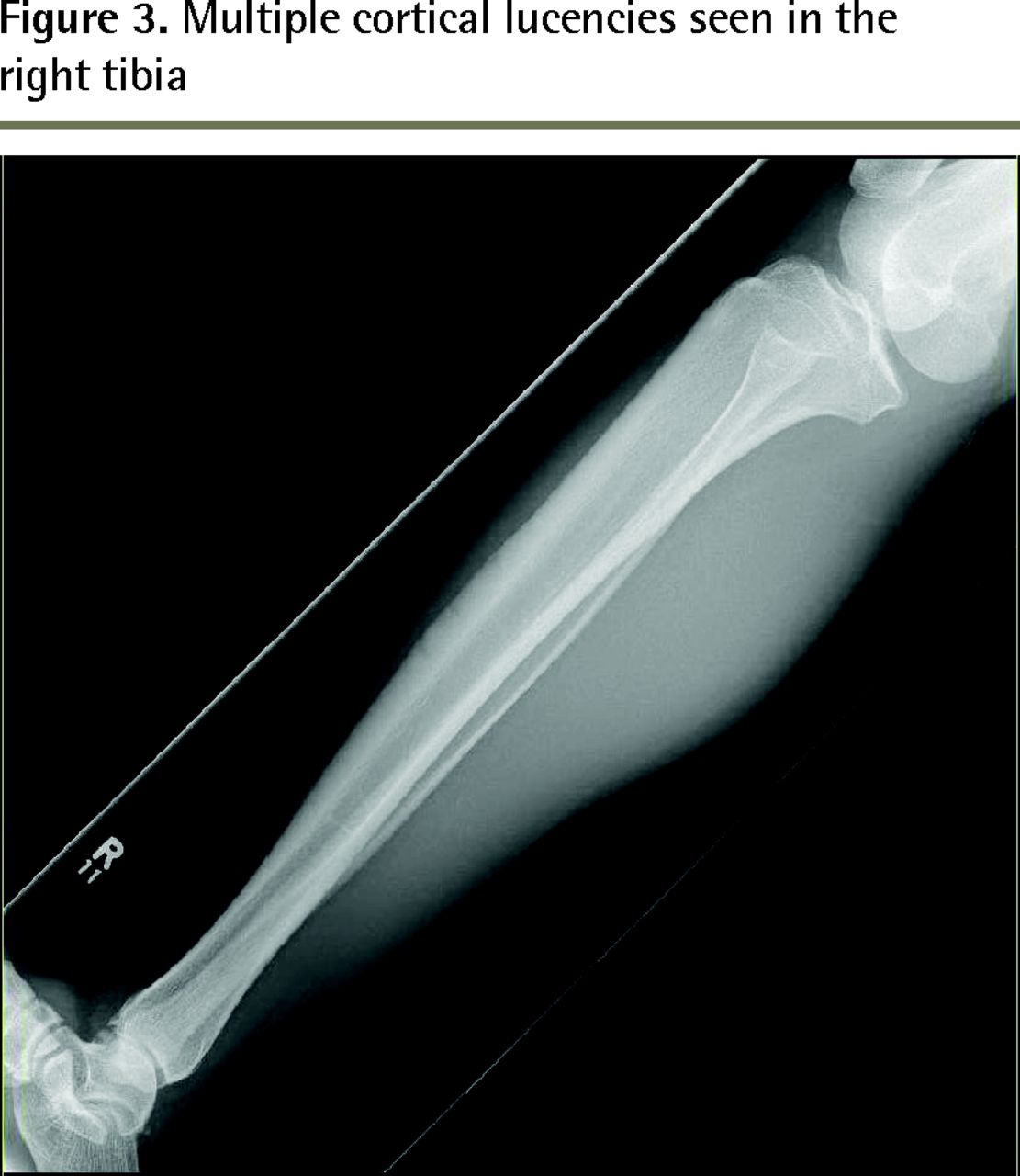
At the time of the orthopedic appointment, J.C. reported clinical improvement, which was corroborated by improvement on radiography. Therefore, surgical management was delayed. Of interest, despite being on strict rest, J.C. presented to our clinic with right shin pain shortly after this, which represented a stress fracture of the contralateral anterior tibia (Figure 3). Low-intensity pulsed ultrasound was initiated immediately under the supervision of the surgeon.

Multiple anterior cortical lucencies seen in the left tibia

“The dreaded black line” signifying nonunion of the stress fracture

Multiple cortical lucencies seen in the right tibia
Discussion
The fundamental approach to treating a stress fracture involves understanding the pathophysiologic forces contributing to the fracture. Initial treatment that includes strict non–weight bearing for up to 6 months with close clinical and radiologic follow-up is often reasonable if the practitioner is comfortable with management. Otherwise, concurrent referral to sports medicine subspecialists or orthopedic surgeons is warranted, especially in refractory stress fractures or if the patient has a history of recurrent low-energy fractures. Further workup, including BMD, biochemistry, and metabolic panel, should also be considered.10 Patel et al provide a concise algorithm for diagnosis and referral that can aid family physicians in managing care of a stress-type injury.11 In our case, J.C. was neither an elite athlete nor had he made any abrupt changes to his training regimen. He had no evident anatomic abnormalities and, similar to the circumstances surrounding the injury, the location of the fracture was atypical. Therefore, workup for underlying pathogenesis was initiated. Of note, this case was managed by an experienced sports and exercise medicine physician and that, along with the fact that J.C.’s clinical symptoms remitted each time he was on strict rest, resulted in a longer period before orthopedics consultation was required.
The patient’s normal BMD, combined with a lack of other end organ involvement, raised the question of whether this case actually represented an incidental fracture in the presence of asymptomatic PHPT rather than a pathologic fracture. The literature suggests that BMD can be preserved for up to the first 8 years of parathyroid disease and might not always be a good predictor of fracture risk in early stages.12 It is generally accepted that severe PHPT increases risk of fracture: epidemiologic studies have shown that the relative fracture risk is increased in untreated PHPT starting as early as 10 years before diagnosis, but some controversy exists around risk in milder disease.13 However, there are few cases in the literature that support the pathologic fracture hypothesis presented in this case. One case in BMJ Case Reports and another in the Pan African Medical Journal described traumatic and stress-type fractures, both in the context of severe PHPT in young men.14,15 Another case in the Journal of Pediatric Orthopaedics discusses an adolescent baseball pitcher with secondary hyperparathyroidism and 2 upper extremity stress fractures.16 None of these cases definitively discuss pathologic stress fractures in young patients secondary to mild or previously asymptomatic PHPT. However, we argue that this case is a first presentation of symptomatic PHPT and that subsequent early definitive management was prudent and warranted. The delayed healing, along with the similar contralateral stress fracture, might further support this conclusion. Furthermore, once the underlying pathology was corrected, conservative measures eventually began to show success. One caveat is that it is difficult to separate the effects of the reversal of metabolic disease from success of ultrasound therapy.
As of the writing of this report, J.C. continues to undergo LIPUS therapy and has demonstrated clinical improvement. He has not yet returned to activity.
Conclusion
Stress fractures are common athletic injuries that can be stratified into low and high risk. Extrinsic and intrinsic factors should be examined to identify the cause of fracture; PHPT is one example of a modifiable intrinsic factor. Management of the stress fracture should begin with routine conservative treatments. However, midtibial anterior cortical stress fractures are at high risk of nonunion and should be treated aggressively from the outset. There are promising treatment methods that can help fracture healing; however, sports medicine or orthopedic consultation should be an early consideration in high-risk cases. Low-intensity pulsed ultrasound therapy can delay the need for surgical intervention.
Notes
EDITOR’S KEY POINTS
Stress fractures are common athletic injuries and primary hyperparathyroidism can increase the risk of fracture.
In this case there was a question about whether the patient’s fractures were indeed pathologic, but the improved healing subsequent to resection of a parathyroid adenoma supports this conclusion.
Referral to a sports and exercise medicine physician or orthopedic surgeon is important to optimize both extrinsic and intrinsic pathophysiologic mechanisms of stress-type injuries. Optimism about low-intensity pulsed ultrasound should remain tempered; however, it is reasonable in the right settings and under the correct supervision.
POINTS DE REPÈRE DU RÉDACTEUR
Les fractures de stress sont des blessures athlétiques courantes et l’hyperparathyroïdie primaire peut augmenter le risque de fracture.
Dans le présent cas, l’origine pathologique des fractures du patient demeurait incertaine, mais l’amélioration de la guérison à la suite de l’ablation d’un adénome parathyroïdien corrobore cette conclusion.
Il importe de demander une consultation en médecine du sport et de l’exercice ou en orthopédie pour optimiser les mécanismes pathologiques tant extrinsèques qu’intrinsèques des blessures du genre fracture de stress. L’optimisme face aux ultrasons pulsés de faible intensité devrait rester modéré; toutefois, c’est une thérapie raisonnable dans le bon contexte et sous une supervision appropriée.
Footnotes
This article has been peer reviewed.
Cet article a fait l’objet d’une révision par des pairs.
Competing interests
None declared
- Copyright© the College of Family Physicians of Canada
References
In this issue


Jump to section
Related Articles
Cited By...
- No citing articles found.