


You are a busy family physician with a practice that includes obstetrics and teaching medical students. Cervical examinations are a routine part of your on-call duties. You have medical trainees with you, and they have often struggled with the cervical examination.
One night on call, your trainee admits a patient in active labour and reports the cervical dilation to be 7 cm. However, she is uncertain about the accuracy of the examination. She has noticed that findings of her cervical assessments can vary from those of staff by as much as 2 to 3 cm. “I’m not certain if it was the cervix that I was feeling,” she reports.
When you arrive on the floor, your assessment reveals cervical dilation of 3 cm. You realize that your trainee likely just examined the posterior fornix and not the cervix. You wonder, How might I better describe my cervical assessment approach so that it is understood by trainees? A review of the literature reveals that no one has addressed this issue previously.
You realize that the crux of the problem is difficulty steering clear of the posterior fornix and recognizing the different textures. You also realize that the texture of tissue in artificial models is not close enough for simulated training.
The solution? You teach your trainee to recognize the appropriate textures by comparing them to the corresponding oral tissues. Using a gloved hand, your trainee examines the following oral tissues:
The sublingual folds and caruncles (Figure 1) approximate the soft, lumpy vaginal mucosa.
The smooth buccal mucosa (Figure 2) mimics the smooth surface of the dilating cervix.
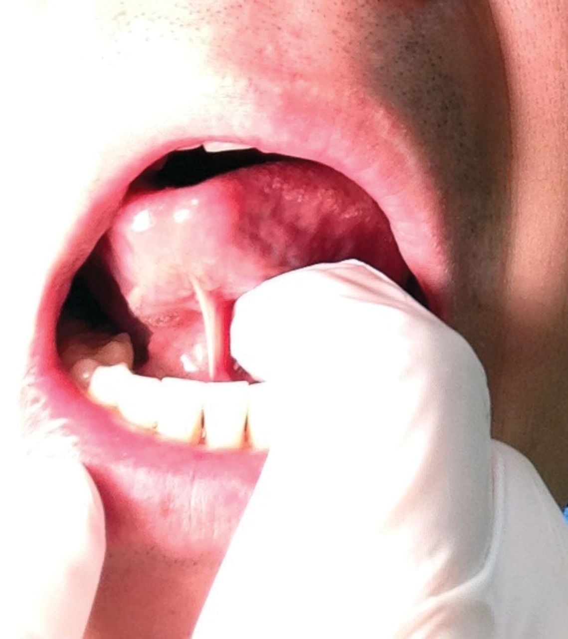
The “sharp” edge formed at the corner of the mouth with a tensed orbicularis oris muscle (Figure 3) feels similar to the cervical os.
Palpating sublingual folds and caruncles to approximate the texture of vaginal mucosa

Palpating the smooth buccal mucosa to approximate the smooth surface of a dilating cervix

Palpating the “sharp” edge formed at the corner of the mouth with a tensed orbicularis oris muscle to approximate the cervical os (edge of cervix)
You teach your trainee an approach to avoid the posterior fornix by maintaining anterior pressure: after inserting your finger 2 cm past the introitus, apply light pressure anteriorly toward the pubic bone. This will prevent you from venturing into the posterior fornix. With anterior pressure, you advance into the vaginal canal while remembering the textures of the equivalent oral tissues, transitioning from sublingual fold, to buccal mucosa, to tensed orbicularis oris muscle (Figure 4).

Cross section of the vagina showing the direction of anterior pressure and insertion of digits (red arrows), with textures noted
With the next patient admission, your trainee applies this new technique. As she now has an approach to the examination and a better understanding of the textures, she successfully determines the cervical dilation.
Notes
We encourage readers to share some of their practice experience: the neat little tricks that solve difficult clinical situations. Praxis articles can be submitted online at http://mc.manuscriptcentral.com/cfp or through the CFP website (www.cfp.ca) under “Authors and Reviewers.”
Footnotes
Competing interests
None declared
- Copyright© the College of Family Physicians of Canada
In this issue


Jump to section
Related Articles
Cited By...
- No citing articles found.