


Orf is one of the most widespread viral diseases worldwide and is caused by Parapoxvirus.1 It can be transmitted to humans from goats, deer, sheep, and cattle. Orf (also known as ecthyma contagiosum) can be transmitted to people who have direct contact with infected animals and it manifests as a erythematous maculopapular lesion. Human orf is usually self-limiting and no specific treatment is needed. Complications are due to misdiagnosis of this infection and, as a result, it being overtreated, specifically by surgical debridement.2
This article describes a case of a 67-year-old man who developed skin lesions on his hands after taking care of goats and sheep on his farm in rural Ontario. Since he retired, he had spent most of his time on his farm away from his home in Toronto, Ont; however, he continued to see his family doctor in Toronto. This case serves as a reminder to urban physicians that it is important to consider zoonotic infection while dealing with skin lesions, as the rate of such infections has increased owing to a combination of demographic changes, increased travel, agricultural practices, and human invasion into animal habitats.3
Case
A 67-year-old Canadian man presented to the emergency department with a complaint of 2 painful blisters on the third and fourth digits of both hands that developed 7 days before the visit. He could not recall any injury or exposure to a similar lesion. The blisters were tender and swollen and their centres became ulcerated and crusted. The patient’s past medical history was unremarkable. He was not taking any medications and had no allergies. He mentioned that he was a retiree from Toronto but recently started to raise goats and sheep to have organic cheese and meat. He also stated that he had not vaccinated his animals for the same reason.
On examination, he was afebrile and the rest of his vital signs were within normal range. The 2 blistered nodules were similar in appearance, about 1.5 cm in diameter and fleshy with ulcerated centres. There was no limitation of range of motion and no associated lymphangitis. One nodule on the right hand had mild surrounding erythema suggestive of a secondary bacterial infection.
The ulcerated centres of the blisters were biopsied, and the tissue was sent for histology, bacterial culture, and sensitivity. The diagnosis of orf was made based on the clinical findings: the history of exposure to goats and sheep, as well as the appearance of the typical cutaneous lesions.
The patient was advised to clean the lesions with antiseptic solution and keep them uncovered. He was discharged with an over-the-counter anti-inflammatory medication for analgesia and 500 mg of cephalexin orally 4 times daily for 7 days for secondary cellulitis. Follow-up 3 days later showed no progression of the lesions. The secondary erythema had resolved. One month later the lesions had completely resolved without residual scarring.
Histologic findings with light microscopy demonstrated acanthotic squamous epithelium with diffuse necrosis of the epidermis and large areas of hemorrhage and crusting with acute inflammatory exudate. The underlying small fragment of papillary dermis showed fibrin deposition and crushed inflammatory cells. The features described above suggested orf (or milkers’ nodules, which is an alternative name for human orf). No viral inclusions were identified. A viral infection could be confirmed by either virologic studies or by ultrastructure examination of the skin biopsy. No further investigations were done, as the nature of the disease was clear and the follow-up visit confirmed the self-limiting condition.
Discussion
Orf is an infection caused by Parapoxvirus that mainly affects goats and sheep. In animals the lesions are localized on lips, nostrils, udders, and toes. The disease is self-limiting and clears without treatment within 3 to 4 weeks. Animals that recover from the infection develop lifelong immunity. A vaccine for animals against orf is available and provides good protection.4
There are limited data on the incidence of human orf, as most orf infections go unreported owing to its self-limiting nature. Also, those who are infected are often familiar with the disease and do not seek medical attention.
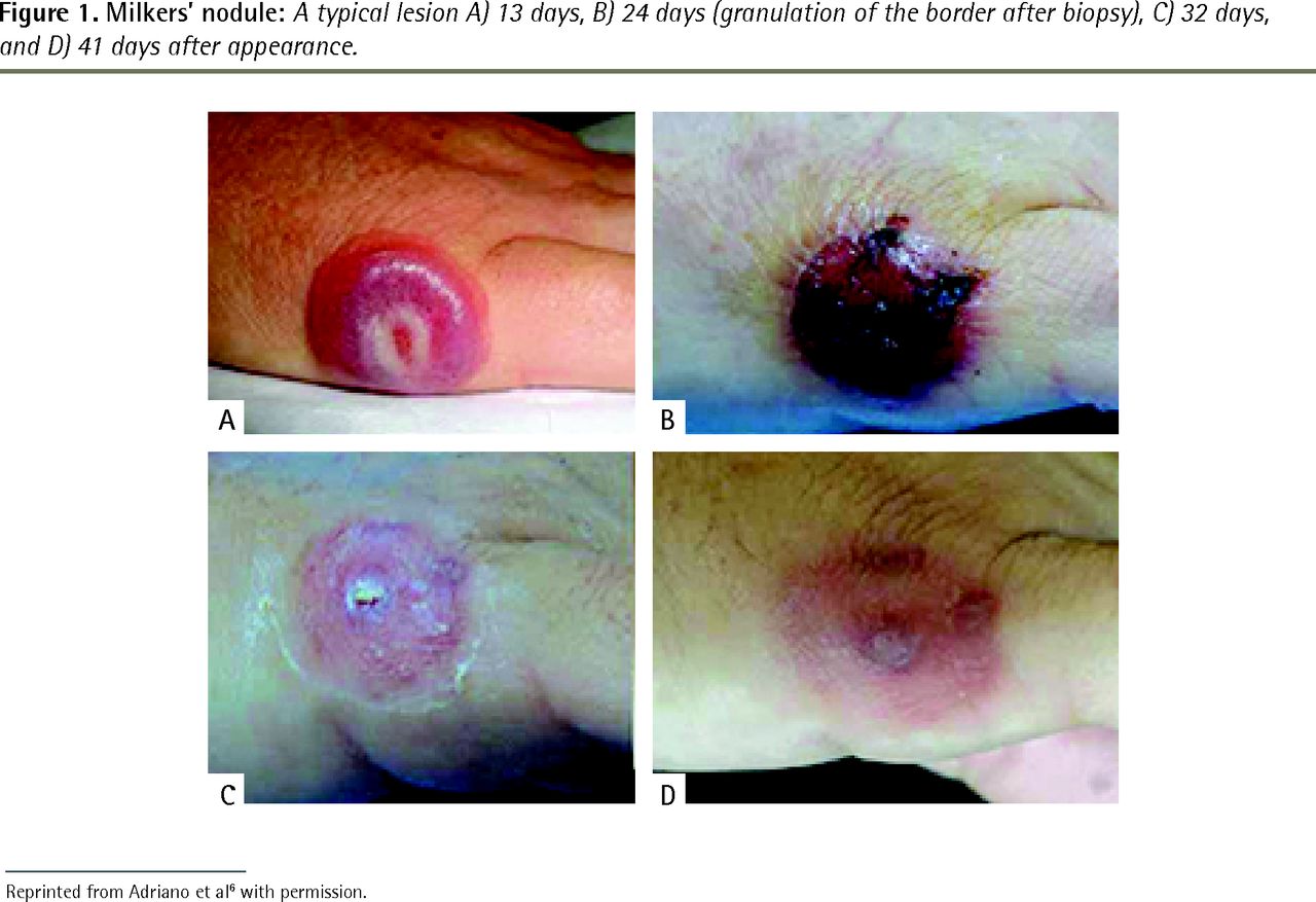
Humans get infected while handling animals, carcasses, or contaminated equipment by direct inoculation through cuts or abrasions. Veterinarians, farmers and their children, wool shearers, and cooks are in the high-risk group for contracting the virus. A lesion appears 3 to 7 days after contact with an infected animal and evolves slowly over the course of 4 to 8 weeks. It starts as a firm red painful papule that expands into a broad, thickened 1- to 3-cm lesion. The centre of the lesion is red and is surrounded by a white, raised ring with erythematous periphery. The crusting occurs and resolves by 3 to 6 weeks with little to no scarring. Recurrent lesions are possible, as the infection does not confer immunity in humans.5 Figure 1 presents the evolution of a typical milkers’ nodule6; milkers’ nodule is also caused by Parapoxvirus and has the same manifestation as orf.

Milkers’ nodule: A typical lesion A) 13 days, B) 24 days (granulation of the border after biopsy), C) 32 days, and D) 41 days after appearance.
Reprinted from Adriano et al6 with permission.
Plain microscopy might detect viral disease. Definitive diagnosis is possible through electron microscopy by demonstrating typical oval viral particles of Parapoxvirus. Polymerase chain reaction is more specific for identifying DNA of the virus in the specimen regardless of the stage of the disease.7
No treatment, including surgical debridement, is necessary, as the disease is self-limiting. The common approach to treat ulcers with surgical debridement is considered unnecessary in cases of human orf because it might cause scarring and prolong the recovery period. The lesion should be kept clean with antiseptic solution to avoid a secondary bacterial infection. Immunocompromised patients need to be treated with topical imiquimod or cidofovir. In the case of a secondary bacterial infection that might present with erythema, purulent secretion, lymphadenitis, or systemic symptoms, antibiotics would be indicated.2
Differential diagnosis of orf includes several diseases such as cutaneous anthrax, pyoderma gangrenosum, and herpetic whitlow.8 Both human orf and cutaneous anthrax can be acquired from sheep and goats. Thus, history alone cannot be helpful in establishing definitive diagnosis. Cutaneous anthrax lesions are fast spreading and ulcerating, and they quickly respond to an appropriate antibiotic. If diagnosis remains unclear, then electron microscopy can confirm a diagnosis of Parapoxvirus infection; only polymerase chain reaction can definitely identify a viral infection such as orf. Pyoderma gangrenosum is most common in patients with underlying systemic disease. It starts as a vesicle or papule on the trunk or limbs, which then enlarges and ulcerates. Herpetic whitlow manifests as a vesicular rash on fingers, often after contact with a vesicle caused by the herpes simplex virus.
Conclusion
Orf is a viral disease that is well known to farmers and veterinarians. Urban physicians should be aware of it while dealing with skin rashes and lesions and take into account patients’ cultural traditions, dietary customs, and migration to rural areas, as these factors put patients at risk of being exposed to the infection. Human orf is a benign infection and its diagnosis can be based on clinical findings to avoid unnecessary expensive testing and invasive treatment.
Acknowledgments
We thank Ms Maya Ginzburg for assistance with editing of the manuscript.
Notes
EDITOR’S KEY POINTS
Orf, which is well known to veterinarians and farmers, is an infection caused by Parapoxvirus that mainly affects goats and sheep. Humans can contract orf through broken skin while handling infected animals or contaminated equipment. Urban physicians should be aware of orf while dealing with skin lesions and take into account patients’ cultural traditions, dietary customs, and migration to rural areas, as these factors put patients at risk of being exposed to the infection.
In the case of human orf, a lesion appears 3 to 7 days after contact with an infected animal and evolves slowly over the course of 4 to 8 weeks.
Human orf is a benign infection and its diagnosis can be based on clinical findings to avoid unnecessary expensive testing and invasive treatment. The disease is self-limiting and clears without treatment within several weeks. Surgical debridement should be avoided because it can lead to scarring and prolong the recovery period. The lesion should be kept clean with antiseptic solution to avoid a secondary bacterial infection.
POINTS DE REPÈRE DU RÉDACTEUR
L’ecthyma, bien connu des vétérinaires et des agriculteurs, est une infection causée par le parapoxvirus qui affecte principalement les chèvres et les moutons. Les humains peuvent contracter l’ecthyma par une lésion de la peau en manipulant des animaux infectés ou de l’équipement contaminé. Les médecins en milieu urbain devraient envisager l’ecthyma en présence de lésions de la peau et prendre en compte les traditions culturelles des patients, leurs coutumes alimentaires et leur migration vers les régions rurales, puisque ces facteurs exposent les patients au risque de contracter cette infection.
Dans le cas de l’ecthyma chez l’humain, une lésion apparaît de 3 à 7 jours après le contact avec un animal infecté et évolue lentement sur une période de 4 à 8 semaines.
L’ecthyma chez l’humain est une infection bénigne, et son diagnostic peut se fonder sur les constatations cliniques pour éviter des analyses coûteuses inutiles et des traitements invasifs. La maladie est spontanément résolutive et disparaît sans traitement après quelques semaines. Il faudrait éviter le débridement chirurgical, parce qu’il pourrait causer des cicatrices et prolonger la période de guérison. Il faut garder la lésion propre au moyen d’une solution antiseptique pour éviter une infection bactérienne secondaire.
Footnotes
This article has been peer reviewed.
Cet article a fait l’objet d’une révision par des pairs. Can Fam Physician 2017;63:769–71
Competing interests
None declared
- Copyright© the College of Family Physicians of Canada
In this issue


Jump to section
Related Articles
Cited By...
- No citing articles found.