
Main menu

  • Home
  • Articles
    • Current
    • Published Ahead of Print
    • Archive
    • Supplemental Issues
    • Collections - French
    • Collections - English
  • Info for
    • Authors & Reviewers
    • Submit a Manuscript
    • Advertisers
    • Careers & Locums
    • Subscribers
    • Permissions
  • About CFP
    • About CFP
    • About the CFPC
    • Editorial Advisory Board
    • Terms of Use
    • Contact Us
  • Feedback
    • Feedback
    • Rapid Responses
    • Most Read
    • Most Cited
    • Email Alerts
  • Blogs
    • Latest Blogs
    • Blog Guidelines
    • Directives pour les blogues
  • Mainpro+ Credits
    • About Mainpro+
    • Member Login
    • Instructions
  • Other Publications
    • http://www.cfpc.ca/Canadianfamilyphysician/
    • https://www.cfpc.ca/Login/
    • Careers and Locums

User menu

  • My alerts

Search

  • Advanced search
The College of Family Physicians of Canada
  • Other Publications
    • http://www.cfpc.ca/Canadianfamilyphysician/
    • https://www.cfpc.ca/Login/
    • Careers and Locums
  • My alerts
The College of Family Physicians of Canada

Advanced Search

  • Home
  • Articles
    • Current
    • Published Ahead of Print
    • Archive
    • Supplemental Issues
    • Collections - French
    • Collections - English
  • Info for
    • Authors & Reviewers
    • Submit a Manuscript
    • Advertisers
    • Careers & Locums
    • Subscribers
    • Permissions
  • About CFP
    • About CFP
    • About the CFPC
    • Editorial Advisory Board
    • Terms of Use
    • Contact Us
  • Feedback
    • Feedback
    • Rapid Responses
    • Most Read
    • Most Cited
    • Email Alerts
  • Blogs
    • Latest Blogs
    • Blog Guidelines
    • Directives pour les blogues
  • Mainpro+ Credits
    • About Mainpro+
    • Member Login
    • Instructions
  • RSS feeds
  • Follow cfp Template on Twitter
OtherCurrent Practice

Answer: Can you identify this condition?

Eiman Nasseri and Anwar Al Hammadi
Canadian Family Physician December 2007, 53 (12) 2123;
Eiman Nasseri
  • Find this author on Google Scholar
  • Find this author on PubMed
  • Search for this author on this site
Anwar Al Hammadi
  • Find this author on Google Scholar
  • Find this author on PubMed
  • Search for this author on this site
  • Article
  • Figures & Data
  • eLetters
  • Info & Metrics
  • PDF
Loading

Answer to Dermacase continued from page 2113

4. Tinea incognita

Dermatophytes are a group of fungi that can infect the skin, hair, and nails because of their ability to use keratin. The organisms colonize keratin tissues, and inflammation is caused by the host response to metabolic by-products. Dermatophyte infections are called ringworm or tinea and are classified by their location on the body. Tinea of the face (excluding the beard area in men) and limbs is called tinea corporis.1

An infection can occur at any age and is more common in warm climates. There is a range of manifestations, and, depending on host immunity and the species of dermatophyte, lesions can vary in size, depth of involvement, and degree of inflammation; however, the most common lesion morphology is annular. Lesions begin as flat scaly spots and develop a raised border that extends in all directions at varying rates. This “advancing” scaly border often contains red papules or vesicles. As the border progresses outward, the central area becomes brown or hypopigmented and less scaly. There might be 1 or more annular lesions, which could be either pruritic or asymptomatic.1

Identifying the typical pattern of inflammation in different body regions and accurately interpreting a potassium hydroxide (KOH) wet-mount preparation of a scale are sufficient to diagnose dermatophyte infections of the skin, as all dermatophytes respond to the same topical and oral agents. However, species identification by culture is ideally warranted for hair and nail infections. Scalp-hair infections in children can originate from an animal that carries a typical species of dermatophyte. The animal can then be traced and treated to prevent future infections. In contrast, nail plates can be infected with nondermatophytes.1

Tinea incognita is the name given to a dermatophyte infection treated with topical or oral corticosteroids, which then loses some of its characteristic features. Corticosteroids might decrease inflammation and give a false impression that the rash is improving, while dermatophytes flourish because of immune suppression.1 Once the treatment is stopped the rash returns but looks different. Scaling at the margin and a well-defined border might be absent, and the rash might have greatly expanded to demonstrate diffuse erythema, telangiectasia, scattered papules, pustules, and hyperpigmentation.2

Diagnosis

Tinea incognita is most often seen on the groin, face, and dorsal aspect of the hand, and is often misdiagnosed as eczema and treated with topical steroids; however, hyphae can still be recognized on a KOH wet-mount preparation.1

To obtain an adequate specimen, steroid medications should be stopped for about 1 week. By then the lesion will again appear scaly. Obtain a sample of scale by holding a No. 15 surgical blade perpendicular to the skin’s surface and drawing it along the active border. Place the fragments of scale on a microscope slide and soak them with a few drops of 10% to 20% KOH solution. Additional stains such as chlorazol fungal stain,3 Swartz-Lamkins fungal stain, or Parker blue ink4 can be used. Apply a coverslip with gentle pressure over the specimen; the slide can be heated over a lighter or Bunsen burner until the preparation darkens but does not boil. The presence of translucent, branching septate hyphae confirms the diagnosis.1

Treatment

Treatment includes the immediate to gradual cessation of all steroid medication, which can lead to transient flaring of the rash, and implementation of an appropriate antifungal regimen.1 Patients should be warned not to reinstitute the use of steroids on their own.

Our patient improved with topical terbinafine cream applied to the ear twice daily for 4 weeks (Figure 1). His case highlights the ease and importance of performing a KOH wet-mount preparation on any scaly lesion and the value of suspecting a dermatophyte fungal infection when lesions persist despite treatment with corticosteroids.

Figure 1
  • Download figure
  • Open in new tab
Figure 1

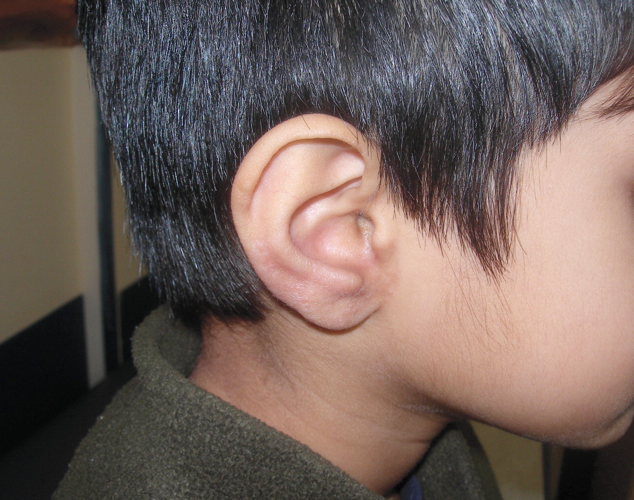
Resolution of the lesion on the right ear after treatment with topical terbinafine cream for 4 weeks

Footnotes

  • Competing interests

    None declared

  • Copyright© the College of Family Physicians of Canada

References

  1. ↵
    1. Habif TP
    1. Habif TP
    . Superficial fungal infections. In: Habif TP, editor. Clinical dermatology: a color guide to diagnosis and therapy. 4. New York, NY: Mosby; 2004. p. 409-26.
  2. ↵
    1. Ive FA,
    2. Marks R
    . Tinea incognito. Br Med J 1968;3(5611):149-52.
    OpenUrlAbstract/FREE Full Text
  3. ↵
    1. Burke WA,
    2. Jones BE
    . A simple stain for rapid office diagnosis of fungus infections of the skin. Arch Dermatol 1984;120(11):1519-20.
    OpenUrlCrossRefPubMed
  4. ↵
    1. Brodell RT,
    2. Helms SE,
    3. Snelson ME
    . Office dermatologic testing: the KOH preparation. Am Fam Physician 1991;43(6):2061-5.
    OpenUrlPubMed
PreviousNext
Back to top

In this issue

Canadian Family Physician: 53 (12)
Canadian Family Physician
Vol. 53, Issue 12
1 Dec 2007
  • Table of Contents
  • About the Cover
  • Index by author
Print
Download PDF
Article Alerts
Sign In to Email Alerts with your Email Address
Email Article

Thank you for your interest in spreading the word on The College of Family Physicians of Canada.

NOTE: We only request your email address so that the person you are recommending the page to knows that you wanted them to see it, and that it is not junk mail. We do not capture any email address.

Enter multiple addresses on separate lines or separate them with commas.
Answer: Can you identify this condition?
(Your Name) has sent you a message from The College of Family Physicians of Canada
(Your Name) thought you would like to see the The College of Family Physicians of Canada web site.
CAPTCHA
This question is for testing whether or not you are a human visitor and to prevent automated spam submissions.
Citation Tools
Answer: Can you identify this condition?
Eiman Nasseri, Anwar Al Hammadi
Canadian Family Physician Dec 2007, 53 (12) 2123;

Citation Manager Formats

  • BibTeX
  • Bookends
  • EasyBib
  • EndNote (tagged)
  • EndNote 8 (xml)
  • Medlars
  • Mendeley
  • Papers
  • RefWorks Tagged
  • Ref Manager
  • RIS
  • Zotero
Respond to this article
Share
Answer: Can you identify this condition?
Eiman Nasseri, Anwar Al Hammadi
Canadian Family Physician Dec 2007, 53 (12) 2123;
del.icio.us logo Digg logo Reddit logo Twitter logo CiteULike logo Facebook logo Google logo Mendeley logo
  • Tweet Widget
  • Facebook Like
  • Google Plus One

Jump to section

  • Article
    • 4. Tinea incognita
    • Footnotes
    • References
  • Figures & Data
  • Info & Metrics
  • eLetters
  • PDF

Related Articles

  • Question: Can you identify this condition?
  • Google Scholar

Cited By...

  • No citing articles found.
  • Google Scholar

More in this TOC Section

Current Practice

  • Scurvy
  • Intraprofessional relationships
  • Question: Can you identify this condition?
Show more Current Practice

Dermacase

  • Question: Can you identify this condition?
  • Answer: Can you identify this condition?
  • Question: Can you identify this condition?
Show more Dermacase

Similar Articles

Navigate

  • Home
  • Current Issue
  • Archive
  • Collections - English
  • Collections - Française

For Authors

  • Authors and Reviewers
  • Submit a Manuscript
  • Permissions
  • Terms of Use

General Information

  • About CFP
  • About the CFPC
  • Advertisers
  • Careers & Locums
  • Editorial Advisory Board
  • Subscribers

Journal Services

  • Email Alerts
  • Twitter
  • RSS Feeds

Copyright © 2022 by The College of Family Physicians of Canada

Powered by HighWire