


Tension pneumothorax is a potentially life-threatening medical emergency that is routinely encountered by family physicians in the course of their busy medical practice. A high index of clinical suspicion, when accompanied by good interpretation of chest radiographs, results in prompt diagnosis and rapid management in these cases. We report a case of right-sided tension pneumothorax that had typical clinical features and classic radiographic findings.
Case
A 43-year-old man presented to his family physician with generalized pain over the right side of his chest following a harsh bout of coughing. The pain rapidly increased in severity over a period of 1 hour and also began radiating to his right arm. Initially, he tolerated the pain, thinking that he might have strained a muscle while coughing, but when it did not abate on resting and appeared to increase in severity, he felt worried. He also began to feel increasingly breathless. On examination, his doctor noticed that he was tachypneic, with a respiratory rate of 32 breaths per minute. His pulse rate was 122 beats per minute and he appeared to be mildly cyanotic. His blood pressure was 92/64 mm Hg. Accessory muscles of respiration were also active. On examination of the chest, the doctor noticed that the right hemithorax appeared to be hyperexpanded and moved less compared with the left. The trachea was deviated to the left and there was a hyperresonant note on percussion over the entire right hemithorax. On auscultation, sounds of air entry were absent over the entire right hemithorax, both anteriorly and posteriorly, while normal vesicular breath sounds were heard over the left side. Vocal resonance was absent on the right side, while it was normal on the left side. The cardiac apex appeared to have shifted outward and was felt in the fifth left intercostal space, 3 cm lateral to the midclavicular line. The patient underwent an urgent chest radiograph. On viewing the chest radiograph, his family physician advised immediate hospitalization.
When the patient arrived at the hospital, he complained of severe right-sided chest pain and appeared acutely breathless. The right hemithorax appeared hyperexpanded compared with the left and did not move with respiration. There was a hyperresonant note on percussion, breath sounds were absent, and vocal resonance could not be elicited over the right side. The chest radiograph (Figure 1) showed a substantially expanded right hemithorax compared with the left. The mediastinum was shifted to the left side. The right side appeared grossly hyperlucent, with a total absence of normal bronchovascular lung markings, which suggested air in the pleural space. There was also considerable widening of the intercostal spaces on the right side, with depression of the right hemidiaphragm, which suggested increasing right intrapleural pressure. In view of the patient’s history, physical examination, and chest radiograph findings, a diagnosis of right-sided tension pneumothorax was made and urgent chest decompression was performed with a needle thoracostomy. An immediate rush of air out of the chest was observed; the patient felt better and air entry improved on the right side. His chest pain abated and his breathing strengthened. An intercostal drainage tube was subsequently inserted. By the fifth day, the right lung had fully expanded and the patient had a good recovery; he was discharged from the hospital on the eighth day. He was advised to refrain from lifting heavy weights and to avoid air travel, and was asked to follow up after 1 week.
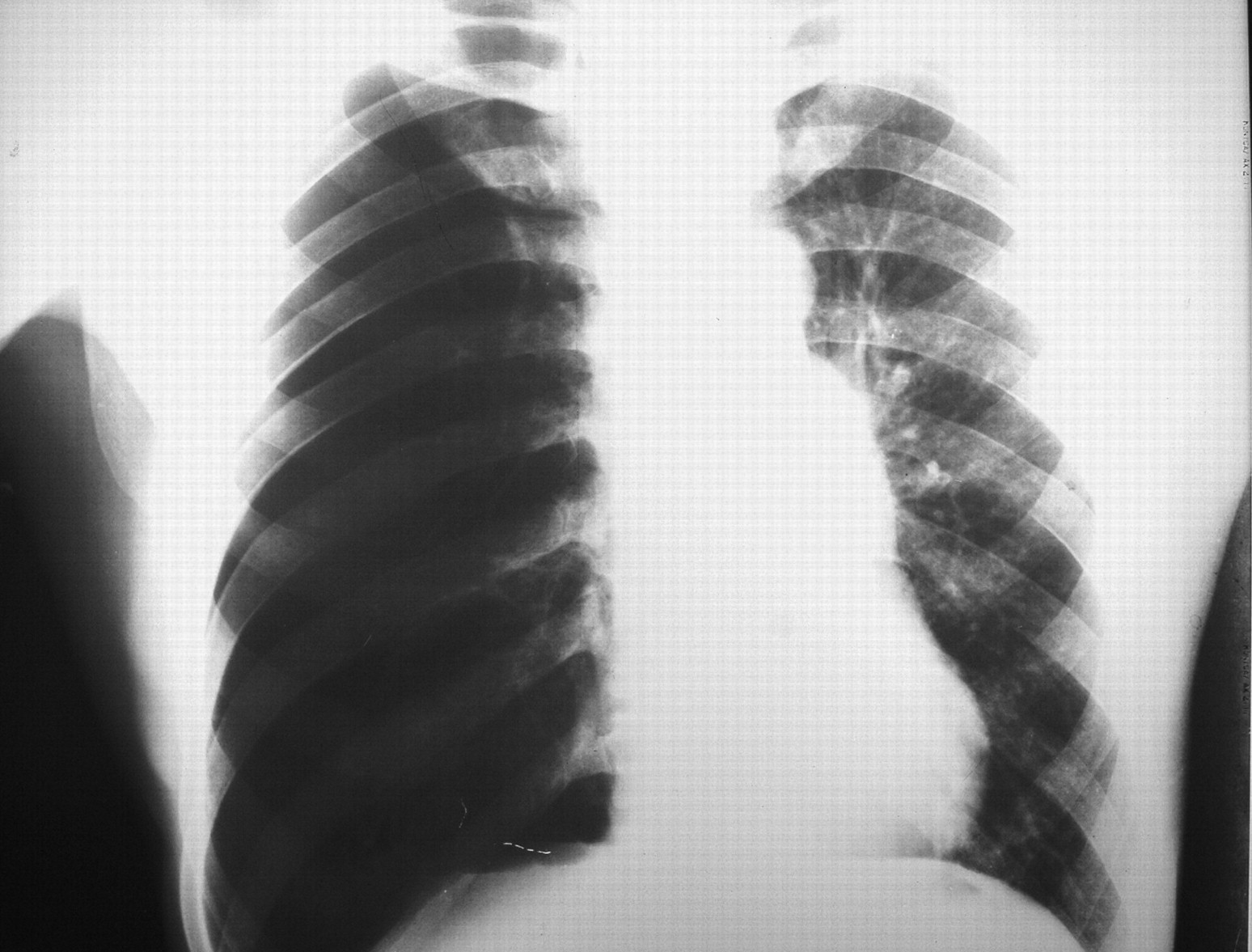
Chest radiograph showing a grossly hyperinflated right hemithorax compared with the left: The right side appears hyperlucent, with absent normal bronchovascular lung markings (suggestive of air in the pleural space), and widened intercostal spaces with depression of the right hemidiaphragm (suggestive of increasing intrapleural pressure); these radiographic findings are suggestive of rapidly increasing tension pneumothorax in a symptomatic and clinically compromised patient.
Discussion
Tension pneumothorax is defined as the presence of air in the pleural cavity that progressively continues to build up. It is due to a lung laceration resulting in a 1-way valve mechanism that allows air to enter the pleural space but not to return. It must be emphasized that tension pneumothorax is a clinical diagnosis. Visual signs might at times be more informative than auscultatory findings in this emergency situation. Clinical signs of tracheal deviation, unilateral hyperexpansion, and hypomobility are classic features that should alert a physician to the possibility of tension pneumothorax when accompanied by increasing breathlessness and unilateral chest pain. Leigh-Smith and Davies1 have thoroughly discussed the importance of these clinical signs in a case report of a patient who developed tension pneumothorax following a severe fall. Others have also pointed out that lateralizing signs, such as hyperresonance and decreased air entry, have been noted to be absent in some advanced states of the disease process.2,3 Werne and Sands4 reported a case of left-sided tension pneumothorax with variable symptoms and signs that masqueraded as an anterior myocardial infarction with characteristic electrocardiographic changes. Chest radiography finally revealed the underlying tension pneumothorax. Teplick and Clark5 suggested that radiologic features of hyperinflation and ipsilateral depression of the hemidiaphragm could be late signs of tension; early signs of tension could include hypoexpansion and hypomobility due to collapse of the lung and pain that limits thoracic wall movements.
Radiologic diagnosis of pneumothorax can be difficult, especially when the pneumothorax is accompanied by chronic obstructive pulmonary disease. A radiologic diagnosis of pneumothorax is made in most cases by identifying the visceral pleural line. However, when pneumothorax is strongly suspected but the visceral pleural line is not identified (possibly obscured by an overlying rib), a chest radiograph in the erect position in full expiration might be required; in an expiratory film, the lung density is increased while the volume of gas in the pleural space remains constant, making it easier to detect pneumothorax.6 Alternatively, a chest radiograph in the lateral decubitus position might be useful because air rises to the highest point and is more clearly visible over the lateral chest wall than over the apex.6,7 In some cases, the only important abnormality might be the deep sulcus sign, in which the usually small space between the chest wall and the diaphragm appears enlarged due to the presence of air, accompanied by a much sharper than normal appearance of the hemidiaphragm.8
Tension pneumothorax, when strongly suspected, requires urgent decompression with needle thoracostomy, followed by intercostal drainage. In an emergency situation, confirmation of the diagnosis with imaging studies might not be possible and it is considered unwise to delay chest decompression in such cases. While reporting the case of a 65-year-old man who had undergone a previous pneumonectomy on the left side and later developed tension pneumothorax on the right side, Watts and Howell9 emphasized that if there is a strong clinical suspicion of tension pneumothorax in a clinically compromised patient, chest decompression must be attempted using needle thoracostomy, if necessary, before radiologic confirmation. Needle thoracostomy is performed in the second intercostal space at the midclavicular line, or 1 to 2 cm lateral to it. The area is first cleaned using povidone-iodine and alcohol before inserting the needle. A large-bore needle (14- or 16-gauge needle) with a catheter is inserted just above the upper border of the third rib. Care should be taken that the needle not be inserted along the lower border of the second rib to avoid damage to the intercostal neurovascular bundle. The needle should be of adequate length (usually more than 4.5 cm) so that it might easily traverse the thickness of the chest wall.10 The needle should be held perpendicularly while it is inserted into the chest wall, and the pressure applied should be well controlled so that the needle is pushed no further once a “give” is felt, which indicates that the parietal pleura has been punctured and the pleural space entered. Once the needle is in the pleural space, a hissing sound will be heard, which indicates the release of air (which is under pressure) from the pleural cavity. The needle is then removed while the catheter is secured and left in place along with a flutter valve.
Needle thoracostomy is usually followed by tube thoracostomy (intercostal drainage), which is the definitive treatment of tension pneumothorax. The tube is usually inserted in an area under the axilla known as the safe triangle. This is done to avoid damage to internal organs. The safe triangle is formed by a horizontal line extending from the nipple and the borders of the latissimus dorsi and pectoralis major muscles. The area is cleaned with povidone-iodine and alcohol and then well anesthetized using a local anesthetic. A small-bore tube (no. 14 French gauge, 4.7-mm diameter) can be inserted using the Seldinger technique.11 In this procedure, the pleural cavity is entered using a sharp, hollow needle called a trocar. A round-tipped guide wire is then inserted through the lumen of the trocar and the trocar is withdrawn. A drainage tube is then passed over the guide wire and the guide wire is withdrawn. The drainage tube is then secured in place and attached either to an underwater seal or a Heimlich valve.
If a large-bore tube (no. 28 French gauge, 9.3-mm diameter) is required, chest tube insertion can be performed by surgically dissecting through the soft tissue down to the rib using a curved hemostat. The intercostal muscles and the parietal pleura are then punctured and a finger is introduced alongside the hemostat. The hemostat is removed and, using a clamp, the intercostal drainage tube is inserted into the pleural cavity alongside the finger. The tube is attached to an underwater seal and secured in place.
Conclusion
Tension pneumothorax is a medical emergency that can be promptly diagnosed if there is a high index of clinical suspicion. General practitioners usually encounter such cases as a consequence of chest trauma. Therefore, they must always be alert to this possibility. In this case, the family physician did well in clinically suspecting pneumothorax and urgently ordering a chest radiograph. This resulted in prompt diagnosis and rapid management.
We believe this is a case of substantial clinical importance because tension pneumothorax is a serious medical emergency that needs to be promptly diagnosed and urgently treated. Moreover, it is commonly encountered by both family physicians and internists in the course of their busy medical practices.
Notes
EDITOR’S KEY POINTS
-
Tension pneumothorax occurs when air progressively builds up in the pleural cavity. It is commonly diagnosed by identifying the visceral pleural line on a chest radiograph.
-
When accompanied by increasing breathlessness and unilateral chest pain, clinical signs of tracheal deviation, unilateral hyperexpansion, and hypomobility should alert the physician to the possibility of tension pneumothorax.
-
Tension pneumothorax requires urgent decompression by needle thoracostomy, followed by intercostal drainage. In an emergency situation, there might not be enough time to confirm the diagnosis with imaging studies.
POINTS DE REPÈRE DU RÉDACTEUR
-
Un pneumothorax sous tension se produit quand l’air s’accumule progressivement dans la cavité pleurale. Il est habituellement diagnostiqué par l’identification de la ligne pleurale viscérale sur une radiographie du thorax.
-
Lorsqu’ils s’accompagnent d’un essoufflement croissant et d’une douleur unilatérale à la poitrine, les signes cliniques de déviation trachéale, d’hyperexpansion unilatérale et d’hypomobilité devraient alerter le médecin de la possibilité d’un pneumothorax sous tension.
-
Un pneumothorax sous tension exige une décompression urgente par thoracostomie à l’aiguille suivie d’un drainage intercostal. Dans une situation d’urgence, il peut ne pas y avoir suffisamment de temps pour confirmer le diagnostic par imagerie.
Footnotes
-
This article has been peer reviewed.
-
Cet article a fait l’objet d’une révision par des pairs.
-
Competing interests
None declared
- Copyright© the College of Family Physicians of Canada
In this issue


Jump to section
Related Articles
Cited By...
- No citing articles found.