


Case
A 22-year-old (gravida 3, para 1, aborta 1) woman was admitted to hospital at 39 weeks and 5 days of gestation with contractions and light blood show (ie, minor bleeding with mucus that is associated with cervical dilation). Shortly after arrival, her membranes ruptured spontaneously for a moderate amount of clear amniotic fluid. Vaginal examination revealed cervical dilation of 5 to 6 cm, cephalic presentation 2 cm above spines, and no untoward events. An initial fetal heart tracing showed normal results, and subsequent intermittent auscultation demonstrated no evidence of fetal distress.
At full dilation the patient had a rapid second stage of labour, lasting only 11 minutes, and delivered a live baby girl weighing 3167 g. There was no evidence of abnormal bleeding. The baby’s Apgar scores were 6 at 1 minute and 8 at 5 minutes. Cord pH was 7.297 with a base excess of −2.5.
The placenta was delivered 11 minutes after the baby, and the following tachycardia-inducing abnormality was noted. Five main vessels traversed the membranes, originating from various sites around the edge of the placenta. A total of 2 veins and 3 arteries left the marginal edge and traversed the sphere enclosed by the membranes. At the polar opposite side of the amniotic sphere, these vessels converged to form a normal umbilical cord with a configuration of 2 arteries and 1 vein. The membrane rupture occurred parallel to, and between, 2 of the vessels (1 artery, 1 vein). Fortunately none of the vessels was injured at the time of the rupture or in the course of delivery. The baby clinically showed no evidence of anemia and no hemoglobin was obtained. Blood transfusion was not required.
The mother’s medical history included antibiotic treatment twice during pregnancy for identified infections (chlamydia at 9 weeks of gestation; abscessed tooth at 19 weeks). This was a spontaneous conception. Her previous pregnancy had ended with a spontaneous vaginal delivery, and there was no record of any placental problems or bleeding.
Routine screening by transabdominal ultrasound at 19 weeks and 4 days of gestation showed an anterior placenta not covering the cervical os. The report made no comment on the umbilical cord or vessels. The baby was noted to be in transverse lie. A repeat transabdominal ultrasound, which was completed at 32 weeks and 4 days of gestation to reassess the baby’s position, showed the baby in cephalic presentation, with the placenta anterior not encroaching on the cervical os. There was no concern identified regarding the umbilical cord.
Discussion
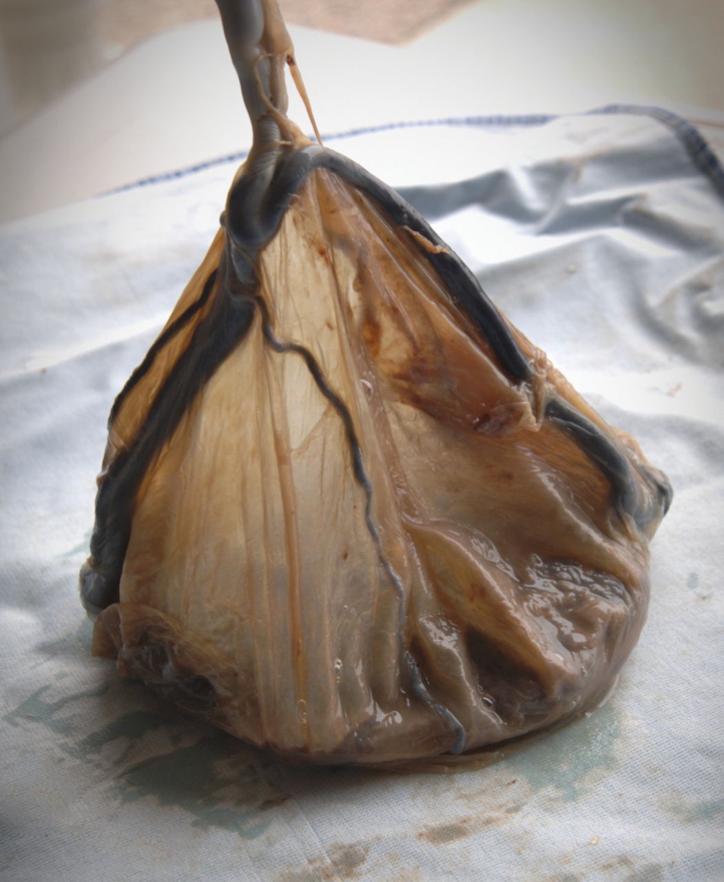
Ruptured vasa previa is a serious and often fatal (to the fetus) obstetric emergency, which was first described in 1801 by Lobstein.1 Before the advent of ultrasound imaging, vasa previa was solely a clinical diagnosis consisting of a triad of ruptured membranes, painless vaginal bleeding (fetal bleeding), and fetal distress or demise. In obstetric terms, vasa previa is defined as “fetal vessels crossing or running in close proximity to the inner cervical os. These vessels course within the membranes (unsupported by the umbilical cord or placental tissue) and are at risk of rupture when the supporting membranes rupture.”1 It is traditionally classified into 2 types. Type 1 is when the vessels run together in front of the cervical os from a site of membranous insertion to the point of cord formation. Type II occurs when bridging vessels between an accessory lobe (succenturiate lobe) and the main body of the placenta (containing the cord) lie in front of the cervix. In the case presented here, 5 separate umbilical vessels left the margin of the placenta at diverse spots and traversed half the circumference of the membranes in different directions before converging to form the umbilical cord. Figures 1 to 4 demonstrate this version of an uncommon placental formation.

Vessels traversing membranes and converging to form cord (formalin-fixed specimen; white balloon to demonstrate shape)

Rupture site and vessels (formalin-fixed specimen; white balloon to demonstrate shape)

Placental margin showing membranous departure of vessels (formalin-fixed specimen)

Membranes showing course of vessels to cord insertion (formalin-fixed specimen)
Velamentous cord insertion is reported in approximately 1% of all pregnancies.1 Vasa previa is reported in 1 out of 1250 to 1 out of 8333 pregnancies.2 Risk factors for vasa previa include low-lying placenta, multi-lobed placenta, velamentous cord insertion, multi-fetal pregnancy, and assisted conception.1 When vasa previa is identified in pregnancy, it is considered an indication for delivery by cesarean section. The survival rate in undiagnosed vasa previa is reported to be only 44%.3
The challenge with this, as with other rare anomalies, is how much investigation should be required for a routine pregnancy in an attempt to identify a rare, but potentially fatal, risk factor. A spate of articles suggests selective prenatal ultrasound screening to detect potential vasa previa.1,3–5 Most of the suggested techniques involve transvaginal ultrasound (a more invasive procedure than transabdominal scanning) to identify paired vessel echoes in the region of the cervix as an attributable characteristic. Where easily identified predisposing risk factors associated with vasa previa exist (ie, multi-fetal pregnancy, low-lying placenta, and assisted conception), such invasive techniques might be warranted. Improved resolution with new ultrasound technology might also be able to include other identifiable placental anomalies such as accessory lobes and velamentous insertion of the umbilical cord. The case presented here would clearly not have met any of the screening criteria. Even if the enhanced screening had been used, the anomaly would likely not have been identified, as all of the vessels in this case traversed the membranes singly (rather than paired). This case reminds us that some of the greatest potential dangers in obstetrics are those we cannot anticipate.
Notes
EDITOR’S KEY POINTS
-
Vasa previa is a serious and often fatal (to the fetus) obstetric emergency.
-
Risk factors for vasa previa include low-lying placenta, multi-lobed placenta, velamentous cord insertion, multi-fetal pregnancy, and assisted conception.
-
For cases in which easily identified predisposing risk factors associated with vasa previa exist, invasive techniques (eg, transvaginal ultrasound) might be warranted in order to identify paired vessel echoes in the region of the cervix. Improved resolution with new ultrasound technology might also be able to identify other placental anomalies such as accessory lobes and velamentous insertion of the umbilical cord that would warrant more aggressive investigation.
-
If enhanced screening had been used in this case, the anomaly would most likely still not have been identified, as all of the vessels traversed the membranes singly rather than in paired fashion. This case reminds us that some of the greatest potential dangers in obstetrics are those we cannot anticipate.
POINTS DE REPÈRE DU RÉDACTEUR
-
Le vasa prævia est une urgence obstétricale sérieuse et souvent fatale (pour le fœtus).
-
Au nombre des facteurs de risque d’un vasa prævia figurent un placenta à insertion basse, un placenta multilobé, une insertion vélamenteuse du cordon, une grossesse multiple et la fécondation assistée.
-
Dans les cas où il existe des facteurs de risque de prédisposition à un vasa prævia facilement indentifiables, des techniques invasives (p. ex. échographie transvaginale) pourraient être justifiées dans le but de capter les ondes de vaisseaux en paires dans la région du col utérin. La meilleure résolution obtenue avec la nouvelle technologie échographique pourrait aussi permettre l’identification d’autres anomalies placentaires, telles que les lobes accessoires et l’insertion vélamenteuse du cordon ombilical, qui exigeraient une investigation plus rigoureuse.
-
Si un meilleur dépistage avait été utilisé dans le cas présent, l’anomalie n’aurait tout de même probablement pas été dépistée car tous les vaisseaux traversaient les membranes comme s’ils étaient uniques plutôt qu’en paires. Ce cas nous rappelle que certains des plus grands dangers potentiels en obstétrique sont ceux que nous ne pouvons pas prévoir.
Footnotes
-
This article has been peer reviewed.
-
Cet article a fait l’objet d’une révision par des pairs.
-
Competing interests
None declared
- Copyright© the College of Family Physicians of Canada


