

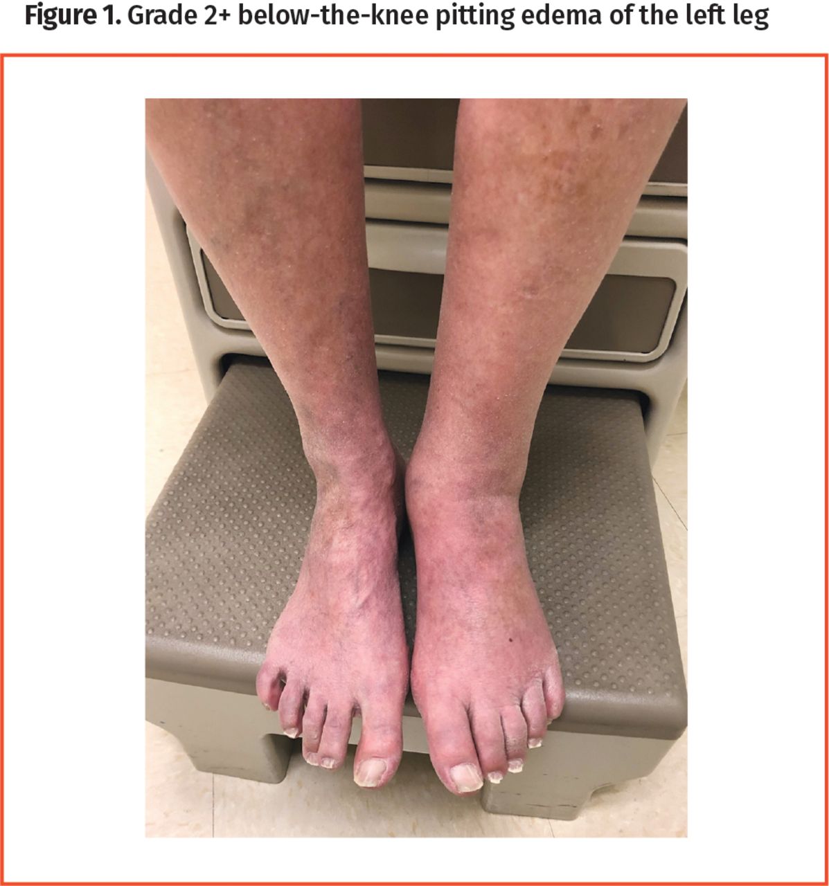
A 76-year-old previously healthy male presented to the clinic with painless swelling in his left leg that he had been experiencing for 1 week. He reported no trauma, travel, recent surgeries, or immobilization and said he did not take any medications on a regular basis. He had no known personal or family history of clots, clotting disorders, or cancers. On physical examination of his left leg, we observed grade 2+ pitting edema below the knee (Figure 1), swelling in the calf (<3 cm when compared with the right calf, and measured 10 cm below the tibial tuberosity), and weak dorsalis pedis and tibialis anterior pulses; the leg and calf were nontender to palpation. We calculated a Wells score of 1 for this patient, but in the absence of a more compelling alternative diagnosis, deep-vein thrombosis (DVT) was at the top of our differential diagnoses list. Accordingly, the patient was scheduled for an urgent ultrasound scan once his blood samples were taken. The ultrasound scan showed occlusive DVT in one of the peroneal veins at the distal aspect of the calf. Later, results of a complete blood count and comprehensive metabolic panel were normal, and a quantitative assay of dimerized plasmin fragment D (D-dimer) showed a concentration of 420 fibrinogen equivalent units (normal range <500 fibrinogen equivalent units).
Grade 2+ below-the-knee pitting edema of the left leg
The American Academy of Family Physicians, the American Society of Hematology, and the American College of Chest Physicians all recommend a stepwise workup for DVT that includes risk stratification, screening laboratory tests, and imaging studies.1-3 A validated prediction rule, most commonly the Wells score, can guide physicians to the appropriate next step.1 While the Wells score was originally conceived for 3-tiered stratification, it is now used to stratify patients into either low or high risk of DVT.4 Patients in the low-risk category (Wells score <2) with a concurrent negative D-dimer test result can be effectively ruled out from having DVT, while those in the high-risk category (Wells score ≥2) should advance straight to imaging, forgoing additional laboratory workup.4 Modern D-dimer assays have reported sensitivities ranging from 95% to 96%, with specificities ranging from 45% to 61% and a negative predictive value (NPV) range from 97% to 99%.5 While highly sensitive, D-dimer assays are imperfect.6 As the pretest probability for a given patient increases, the NPV of the D-dimer assay decreases and it becomes a less reliable tool for ruling out DVT.4 A Wells score of less than 2 yields a D-dimer test with an NPV of 99.1% (95% CI 96.7% to 99.9%); however, with a Wells score of 2 or greater, the NPV of the test drops to 89.0% (95% CI 80.7% to 94.6%).7 As in our patient, the high pretest probability rendered the D-dimer test’s NPV less reliable and ultimately yielded a false-negative result. This relationship between clinical pretest probability and negative or positive predictive values is applicable to all screening studies and is an area for potential misstep. Discerning physicians should keep this relationship in mind when their clinical suspicions differ from the results of a screening laboratory test.
Footnotes
Competing interests
None declared
This article has been peer reviewed.
- Copyright© 2022 the College of Family Physicians of Canada
References
- 1.
- 2.
- 3.
- 4.
- 5.
- 6.
- 7.